The
human body is a slippery surface upon which discourses of race, class, gender,
and sexuality are mediated, and thus it is a contested scientific, political,
ethical, cultural, economic, and social site. Since human subjectivity and
identity are linked to the changing perceptions of vision and visualization, we
make and remake our visual experiences of the world within these different
contexts. In diagnostic imaging, the areas of visualization, medicine, and
technology come together. Using the term “divining” synonymously with
“diagnosing,” the exhibition title Divining Fragments: Reconciling the Body
style=’font-size:8.0pt;font-family:Verdana;color:black’> refers to the history
of diagnostics, from prognosticating over the internal organs of animals and
ill gotten human specimens to visualizing the unseeable through dissection,
microscopy, sonograms, x-rays, CAT, MRI and PET scans, including alternative
techniques like phrenology, Kirlian and aura photography, as well as total body
scanning from military applications.
Confronted
with a public presentation of our own private exposed anatomy we experience the
loss of boundaries and control, evoking issues of physicality, vulnerability,
and mortality. Through the imaging and re-imagining of the human body these
visual technologies have a profound impact on human self-understanding and
behavior, often through implications outside of clinical application, and bring
the relationships between health and knowledge under essential scrutiny,
questioning the way that meaning is negotiated. The manufacturers of these
machines would have us believe that their technologies produce unbiased images
that reveal truths about an individual’s condition, but discrepancies exist
between “machine vision” and “human vision.” Much of the psychophysical data
used in the past to engineer high-performance networked imaging systems is not
consistent with the current knowledge of the human nervous system, and
compensating enhancements of the image risk misinterpretation and the
introduction of artifacts. The designs of information transmitting, storing and
processing devices need a better fit between opto-electronics and human nervous
systems.
Historically,
the partial or fragmented image suggested grief and nostalgia for the loss of a
vanished totality and a utopian wholeness. In diagnostic imaging the body is examined in detail,
piecemeal and irreconciled, described in terms of “cuts” and “slices.” The
body’s pieces, viewed as relics and synecdoches, constitute deconstructed
images of humans and problematize issues of creation and re-creation, existence
and mortality, integration and dissolution, especially when the images of the
dematerialized body are solely transduced from digital code, existing as pure
information.
Anatomical
dissection has traditionally been the basis of studying the human body in
health and disease despite the ban on human dissection that existed
intermittently throughout the history of medicine. We acknowledge that
DaVinci’s detailed anatomical studies were done covertly, paradoxically at a
time when art and science were more closely aligned than today. When Andreas
Vesalius published “De Humani Corporis Fabrica” in 1543, it had the same
comprehensive impact that modern medical imaging technology had on our view of
the human body: it changed human consciousness, and created new anatomists.
Mark
Kessell’s prints of a sagitally-sectioned head, “The Intersection of Self”
(2003) and a pile of flayed, dissected arms, “Continuing to Act” (2003) refer
to the pre-history of diagnostic imaging when the study of gross anatomy with
its skills of dissection and direct visualization of the skeleton, organs and
tissues was one of the primary tools for training physicians in the detection
of disease and normal variants. An amazing find by vintage photography dealer
David Winter was a series of photograms, which turned out to be transparent
sections of a human body from a New York hospital in the 1920-1930s. These
images resemble but obviously predate CAT scans and were likely created from
cryosections, and they are unique in the way they show both bone and soft
tissue in exquisite detail, especially muscles bundled in fascia. In the
paramedial sagittal view, the headless body is abstracted, marked by the
dramatic dark diagonal stripes of the dense ribs.
When
the field of medicine was transformed from a primarily tactile and aural
diagnostic practice into a visual practice by the introduction of roentgen
rays, the medical body collided with the social body. This was in the late
Victorian era when even the legs of furniture were being covered, and protests
were launched about the indecency of this new viewing of the human body. The
x-ray is a pivotal site of the exploration of medicine’s visual knowledge and
power. Before their full effects were discovered, x-rays were used in the
treatment of acne and fluoroscopes were used in stores as an aid to fitting
shoes; and, of course, this put people at increased risk for injury, mutations,
and cancer from radiation exposure. X-rays are part of the treatment regimen
for cancer, but they were used for involuntary sterilization in by the Nazis,
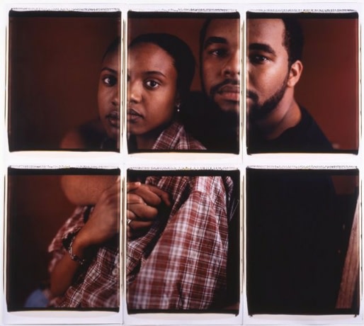
and were a basis for the military “death ray.” One of the first uses of medical
imaging in contemporary art appeared in Rauschenberg’s Booster
style=’font-size:8.0pt;font-family:Verdana;color:black’> (1967), and it
consists of five separate x-rays of the artist’s body reassembled in a six foot
art piece. David Webster is an artist whose work is devoted to the physiologic
and anatomic, including sculptures, photographs, and paintings depicting
histology, disease, and dysfunction. His series in the exhibition are those of
mudras, hand positions of the Buddha that constitute a highly stylized form of
gestural communication. Of those depicted in his lightbox piece Six Sacred
Positions of Buddha (1995) the Dharmachakra mudra denotes the setting of the wheel of
the teaching of the dharma into motion. Webster’s x-ray images using his own
hands in mudras are hauntingly poignant, for they penetrate his own physicality
and vulnerability. As a result of
an illness that required her to have multiple x-rays, Kuni� Sugiura was
inspired to create her Rack (1996) series consisting of photograms
assembled from collected x-rays of anonymous patients. Intrigued by her
unfamiliarity with the people whose images she was handling, she was
additionally inspired by the connection between the medical assessment of their
inner parts and the inner aspects of her own artistic expression. Created in
isolation in the darkroom, these photograms provided her with a rewarding link
to society at large and a satisfying means of recycling these discarded images.
Postcard-sized photograms made from roentgenograms of skulls, chests, and
spines are mounted in a found postcard holder, tethered tensely between the
floor and ceiling at roughly anatomical position.
The
sonogram produces an image on the basis of sound waves reflected back to a
source as a function of density and lucency of organs, tissues, and fluid, and
has found wide application in the assessment of fetuses and pregnancies. Steve
Miller created L’Origine du
Monde
(1994), a large silkscreen painting of a sonogram of twin girls with a
reference to Courbet’s infamous work, bracketed by a radar-like sweep of paint
above, and an EKG tracing below. The artist is deeply invested in the fields of
science and technology, and his works draw from iconic images, cartography, and
the medical body to address the ways that identity is constructed.
Taking
cartography further are the pioneering works of Lilla LoCurto and William
Outcault. Like phrenology yet unlike medical diagnostic imaging devices, these
images are derived only from the surface of the artists’ bodies despite their
resemblance to the “cuts” of internal scanning technologies. Their image-making
process involves total body scans from military application modified by
programs developed in conjunction with a computer scientist and two
mathematicians to map the volumetric body in 2-D, the same goal of simultaneity
sought by the Cubist artists. The work in the exhibition Storyboard#4-5
style=’font-size:8.0pt;font-family:Verdana;color:black’> (2003) is a still
taken from a video that depicts the same imaging technology, where the angle of
the “slice” is programmed to change, resulting in an astounding violently
poetic calligraphic image, just recognizable as the sectioned and unraveled
bodies of the artists.
Advanced
medical imaging technologies came into clinical use in successive decades: CAT
in the 70s, MRI in the 80s, and PET in the 90s. Unlike CAT scans that rely on
the summation of x-ray images and PET scans that rely on the decay of an
injected radioactive pharmaceutical, MRI does not involve radiation, but
instead uses a powerful magnet and the spin of hydrogen atoms in the body’s
water to generate images. It is astonishing to think of MRI and PET scans as
the body’s way of illuminating
itself from within through subatomic particles. The online Visible Human
Project,
sponsored by the National Library of Medicine, imaged the bodies of an executed
murderer and a female body using the full array of medical imaging technologies
including transverse CT, MR, and cryosection images. The male was sectioned at
one millimeter intervals, the female at one third of a millimeter intervals.
The long-term goal of the project is to produce a system of knowledge structure
that will transparently link visual knowledge forms to symbolic knowledge
formats such as the names of body parts. In his untitled 2002 piece, Patrick
Martinez has taken the successive slices from the project and placed them in
rapid sequence to create images of expanding and contracting abstractions,
giving one the mesmerizing sensation of traveling through the body at great
speed.
Justine
Cooper’s spectrally elegant sculpture Reach
style=’font-size:8.0pt;font-family:Verdana;color:black’> (2000) consists of
planar sections of her hands and forearms from MRI data printed on stacked
glass sheets re assembled in 3-D that impel the viewer to move around in space
to reconcile the pieces back into a coherent whole, mirroring the translation
and deciphering processes of MRI while making the interstitial areas the
viewer’s spaces.
The
Cartesian split results from the collision of paradigms between faith and
empiricism, logic and intuition; and should be remembered when society is
willing to abdicate rationality and responsibility to science and technology in
the creation of machines that we are led to believe will tell us the truth
about ourselves. One must recall that the original information produced by MRI
is numerical, not visual, and that the process of reassembling the original
body image can be a conflicted re-presentation. Metal in or on the body,
breathing, and even the movement of blood can produce artifacts termed “cross
talk” that appear as white dots created when the MRI “slices” are too close
together. The very existence of these artifacts refute the notion of MRI making
the body transparent and the image equivalent to the body since the production
process involves the designation of twenty parameters, each of which affects
how the final image appears and what it does and does not show. MRI images,
reconstructed from pure information, should also be understood as knowledge
that is influenced by social context and human decisions and they should not be
separated from the discussions of the people and institutions that construct
and control them. That there are dire consequences of equating photos with the
real have been pointed out by cultural critics John Berger and Susan Sontag.
Medical images circulate similarly within this belief system and are also often
thought to be equivalent to the bodies represented within them. Realizing that
MRI images are only re presentations and partial truths empowers us to
recognize the political, social, and economic factors that affect the
interpretation of these images.
Warren
Neidich’s indexical renderings of heads resemble CAT scans, but in actuality
are photographs of light “paintings” depicting heads in profile that were part
of his project studying prosopagnosia, the inability to recognize faces,
including one’s own. Robert #2 is from the Blanqui’s Cosmology series
(1999-2002) that reminded the artist of phrenology, a respected hard science in
the 19th century that analyzed character through the overall shape of and
protuberances on the skull. When the same outlines were noted to resemble ring
nebulae and other cosmologic references, Neidich’s natural scientific curiosity
and interest in systems formulated this creative bifid reading of his work.
At
the other end of the magnifying lens is Jeff Wyckoff, a cancer and AIDS
research scientist whose position allows him to work at the forefront of new
technology. His piece Alpha-Omega
style=’font-size:8.0pt;font-family:Verdana;color:black’> (2003) presents a new
view of cardiac and tumor tissues through multi photon microscopy that works by
sending a high wavelength infrared laser into a standard light microscope. When
two beams join at the focal plane, the sum of the wavelengths creates light at
normal light microscope wavelengths with the advantage of deeper penetration
and less phototoxicity. With the imagery, he presents a menu of accompanying
music created by four different composers in varying styles.
In
the 1930s the Russian technician Semyon D. Kirlian discovered a means of
showing film imprints of electromagnetic energy as unique fields of color in
living organisms. These photographs may have first been applied as diagnostic
tools or as aids to behavioral modification. Chrysanne Stathacos has traveled
widely to record the results of both psychic and somatic phenomena through
biofeedback plates onto Polaroid portraits of mystics and sadhus that show surrounding clouds of brilliant
varying colors but the artist leaves all interpretations open-ended.
These
deployments of medical imaging pictures by contemporary visual artists reflect
the innovative and alternative perspectives that art often offers to science
with its pursuit of material truths, while acknowledging that both art and
science are investigated by social beings within social contexts. |